First for me, while categorizing. I don’t think I’ve seen this before in such quantity…
What are the bright and white, irregularly-shaped splotches in this video?
Are they large vessels clogged with stalls?
First for me, while categorizing. I don’t think I’ve seen this before in such quantity…
What are the bright and white, irregularly-shaped splotches in this video?
Are they large vessels clogged with stalls?
Thank you for the question @Plenum! I am not sure myself, but I asked our biomedical researchers – let’s see what they say!
Hi again!
Nozomi (@nn62) just responded about this, and looks like it’s not a vessel at all!
This is not a blood vessel, but a cell that has taken up the dye that labels the blood vessels. Overtime, some of the dye in the blood vessels will leak into the surrounding tissue. There are several classes of cell including macrophages that phagocytose or “eat” debris, “cleaning” the tissues. This is likely what is happening here. This is a cell that has gathered up and sequestered the leaked dye.
It also seems to be happening in the dura tissue which should be masked/removed in the preprocessing of the movies, but algorithms are not smart enough to get all of them yet! 
 They are planning to look into this – find out why these surface vessels got in there.
So, with Nozomi’s reply, its the macrophages that gradually absorb the dye, thus producing the splotchy areas next to the vessel walls.
To further this topic, we have a note from Egle regarding the SIZE of the macrophages:
"Yes, macrophages are the largest blood cells I think!
"Here’s some pics for scale:
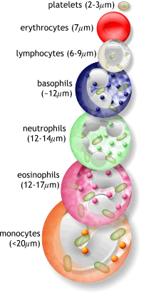
(monocytes are precursors for macrophages)
Those above are human cells – a human macrophage is about 21 micrometers, while a human capillary is about 5-10 micrometers. Those proportions are probably similar in mice too!
Okay-y-y… Well, for me, two take-aways… First, I’m truly surprised at the size of the macrophages., and want to read up on what other sources have to say about the cell. Second, the algorithm, still not perfect, blacks portions of the video, but has not learned to black out the macrophages. This explains the blacked out portions we’ve seen in the last few weeks.
“Mystery of the White Splotch” seems solved… Dye absorbed by Macrophages.
Might take a look at this video at the very beginning… Just my guess, but they appear to be cells (macrophages?) residing on the vessels. If so, this is at a much lower resolution.
OK, just to clarify then, this particular best-fit “vessel” is really not a stall. Correct? Now if the algorithm would just stop featuring them. 
Right, not a stall.
Lol yeah it would be great if the algorithm got it’s marbles together!  (Hoping the lab will deal with this in due course…)
Okay, the latest on the “white splotches” which also include the small white balls seen in an earlier video:
Nozomi (the biomed specialist) replied the following…
“To be honest, I don’t know what these are. Some of the bright deposits look to be in the same position where we see some categories of macrophages, but there are quite a few of them. We would have to do some additional experiments to try to figure this out. One of the challenges of the live imaging with two-photon microscopy is that we only see what we label, or occasionally, what generates fluorescent molecules, like what is happening in these images. To get more information about the cells and structures that we see with in vivo imaging, we will extract the brains after the last imaging session, cut the brain tissue into thin slices and mount the tissue sections onto microscope slides. Then we can apply additional labels that bind to specific cell types and proteins to see other cells that were not visible before.”

 I will ask if those experiments are actually planned or hypothetical
Thanks, Seplute and Nozomi!!
Plenum
Our current dataset seems to contain a lot of the mystery “white splotches” that catcher Plenum first alerted us to in June 2019.
For background, you can scroll to the top of this discussion topic. (I’m not sure the old links will still work.) Many of our current catchers might be confused by movies that the algorithm has selected for annotation, even though there is no real vessel present. Per the old discussion above, these might or might not be macrophages. Sometimes they can look like stalled vessels, but usually are just “unknown” patchy structures.
I’ll try to paste in a couple examples from our current dataset later. The important thing to remember when you see these usually undefined structures is to simply click the flag and then mark them as flowing.
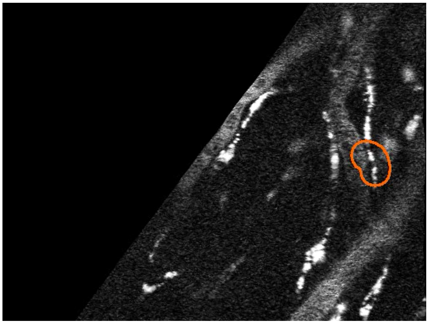
Edit 1 - OK, if it works, here’s example one:
At least the white dots are somewhat aligned, but there is no apparent vessel entering or leaving the orange outline - highly suspect.
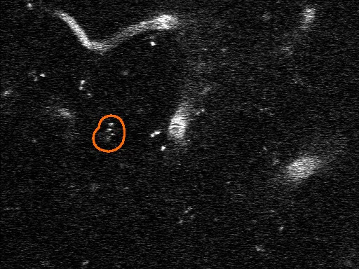
Edit 2 - Hey, that worked! Example two is similar
OK, that should give you the idea, but I’ll look for another example and see if I can link you to the whole image stack (movie) - I need the practice/training. ![]()
![]()
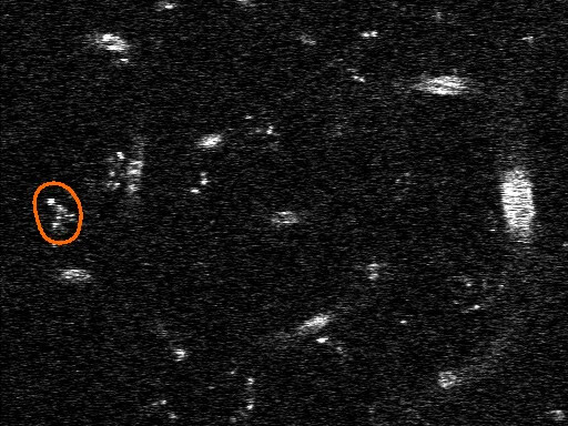
Edit 3 - Here goes, if I guessed correctly, example three:
Whoa, that worked - there’s hope for me yet! Now, if you mark/marked this a stall, I don’t blame you. There appears to be a pattern of dots that kind of lead into and out of the outline, but no vessel! The macrophages tend to accumulate along vessel walls - but what to do with this? Was there a vessel that decomposed? Got abducted by aliens? Got me. Until we get more expert guidance, I’d flag it.
Please, with your indulgence, one more example as better ones keep popping up:
Here we have some tempting, brilliant white splotches, but only for a few frames, and they go no where. Another one to flag!