What is the difference between a stall, and a blood clot? Would blood thinners prevents stalls in much the same way that they are used to prevent blood clots?
Hello, @MikeLandau,
Excellent question!
I will take a stab at this one and then see what our bio experts, @Chris_Schaffer or @nn62, might have to say…
A clot is an obstruction that results from blood cells clumping together. A stall results from inflammation on the inside of a tiny blood vessel (capillary). So even though they can both obstruct blood flow in a vessel, they have different causes (and, therefore, different treatments).
Most of the capillaries in Stall Catchers are only wide enough to allow blood cells to pass through single-file. If a blood vessel becomes inflamed, that basically means a little chemical “hook” appears on the inside of the vessel and snags the next blood cell that happens to come by. Once that blood cell has been caught and held in place, then none of the other blood cells can pass by. They are not stuck together, they are just held up in a traffic jam.
One interesting point about the stalls that we see in Alzheimer’s is that they are very transient - meaning they only last about 10 seconds and then the blood starts flowing again. But as soon as one resolves, another one appears somewhere else. So when we talk about the number of stalled vessels, we are really saying, “what percent of the vessels are stalled, on average, at any given moment?”
Hope that helps, and let’s see if @Chris_Schaffer or @nn62 have anything to add (or subtract!) from what I said.
Best,
@pietro
Good job, Pietro. Just adding a bit more - There were several possible causes of stalled capillaries. As Pietro said, one is a clot. This would come from the same process that you see when you cut your hand and a scab forms. That scab has platelets as well as blood cells. There are other diseases in which we also see stalled capillaries in the brain. For example, in mice that mimic the disease polycythemia vera (patient has too many blood cells), a large number the stalled capillaries are also found in the brain, but these are caused by clots. In the Alzheimer mice, we have observed that most of the stalled capillaries have a particular kind of white blood cell in them - a neutrophil. This is really surprising because neutrophils are the cells that usually react to the invasion of bacteria, but bacterial infections are not associated with Alzheimer’s disease.
Because the stalls we are seeing here are not blood clots, blood thinners like aspirin and warfarin likely will not have a strong effect. In future studies, we are targeting compounds that affect neutrophils more directly.
Thank you for your reply. There is something that you said that I’m still confused about. You said, that there are several possible causes of stalled capillaries, and one of them is a clot. Are you saying that both blood clots, and the types of blockages that we see in the mouse blood vessels in the stall catchers game are types of stalls? That’s what your statement seems to be saying, yet from what Pietro wrote, I got the impression that blood clots, and stalls are two different things.
It is interesting to me that you say that aspirin does not have a strong effect because while it thins the blood, aspirin also reduces inflammation, at least it reduces the inflammation associated with certain types of arthritis. If the stalls that we are seeing in the mouse blood vessels are caused by inflammation of the blood vessels, I would think that aspirin might have some effect. Maybe the aspirin isn’t having any effect because the dosage is not large enough. I am under the impression that when people use aspirin as a blood thinner, they don’t take very much, whereas when people use aspirin to treat arthritis, they take considerably more. For this reason, I would say that if one is treating inflammation of the blood vessels using aspirin, the dosage should be considerably more than when they are using it as a blood thinner. This assumes of course that the inflammation that causes the stalls in the blood vessels is the same as other types of inflammation seen throughout the body, such as arthritis. I don’t know if all types of inflammation arise from the same causes or not. If they don’t, the ideas discussed in my statements above might not make much sense.
Hi @MikeLandau,
Yes - I think your interpretation is correct.
I have probably created some confusion here. A “stall” simply means that blood isn’t flowing in a vessel. I was probably conflating cause and effect in my explanation, because in Stall Catchers, most of these are caused by the same thing: “inflammation” (which I’ll address in your other post). To be clear, both inflammation and clots are two different potential causes of blood flow disruption in a vessel. Sorry for not having been clearer about this in my original explanation. @nn62 may have something to add to this 
Does that help resolve the confusion?
Best,
Pietro
Dear @MikeLandau,
You are full of excellent observations and questions!
As I am not a biologist, I will defer more vigorously to @Chris_Schaffer or @nn62 on this question. However, I think you are on the right track to answering your own question - that is, inflammation is manifests in many ways, and the type of inflammation seen in arthritis, for example, may have very different immunological characteristics and molecular pathways than inflammatory effects observed in brain capillaries.
Inflammation is basically an immune response. Different kinds of inflammation, I believe, may also have different secondary effects. For example, the most commonly associated symptoms of pain, heat, and swelling are caused by some but not all types of inflammation. Inflammation of brain capillaries has a very specific manifestation, the description of which I will leave to the biomedical experts 
All best,
Pietro
To be clear, we have not yet tested asprin specifically, but that may be a good candidate in our upcoming studies. Inflammation is a very complex phenomena. For example in wound healing, different type of cells get recruited to the wound at various times and play distinct roles. There are several negative clinical studies about anti-inflamatories in AD. My suspicion is that we need to target a specific aspect of this very complex pathway rather than broadly inhibit the whole thing.
Okay, thanks! I think I understand. So whether a blood flow disruption is caused by inflammation, or a blood clot it can be referred to as a stall? I guess my confusion stems from this idea I have that a blood clot is a rather long-lasting disruption of blood flow, whereas a stall is a much briefer disruption in blood flow. I think I got that idea from watching the movies in the game because while the stalls cause a disruption, they still can be seen moving through the blood vessel, so they appear to be quite brief in nature.
Okay, thanks! I wanted to ask you another question. Earlier, you said that you have observed neutrophils in the stalls capillaries. If there are no bacteria present to produce this reaction, do you think this could be an indication of some kind of autoimmune response? It seems recently that many diseases are being found to have some kind of autoimmune response as their underlying cause. Has anyone considered whether or not Alzheimer’s could be due to the immune system attacking neurons, and also possibly attacking the lining of these blood vessels, and causing the inflammation?
Aha! Another good question and a great opportunity for a point of clarification…
The dark spots in the blood vessels are blood cells. When they are moving, the vessel is not stuck. But when, over the course of a movie (moving the slider back and forth), a dark spot never goes away, that is considered a stall.
So when you wrote “they can still be seen moving through the vessel” - in that case, I would score that vessel as “flowing” rather than stalled. Put another way, it is impossible to tell if a vessel is flowing or stalled from a still image. The only way to tell, is to see what happens to those dark spots as you move the slider.
The transience of stalls (how briefly they last) tends to be around 10-15 seconds. So while it is possible for vessel movies to capture that exact moment when a stall releases, in the vast majority of cases, it is either flowing or stalled and not both.
Does that help?
Thanks for this great discussion thread!
Best,
Pietro
Okay, here’s what I don’t understand. As I move the slider, the light source grows in intensity, and then fades away. All the spots appear to move at one point or another, it just depends how bright the light is. Why does the light source constantly change in intensity? It’s like trying to find something in your darkened closet, but there’s only one lightbulb that is swinging back and forth constantly illuminating one point, or another. Does a stall mean seeing three dark spots in quick succession? That’s what the tutorial movie appears to indicate. If I see one or two dark spots, but not three is that still a stall? One time, there was only one dark spot and the expert still called it a stall.
Also, what what is the orange circle all about? Why are we only supposed to be looking for stalls in the orange circle? A couple times, I saw a stall, but it disappeared before it reached the orange circle, so I guess in that case it didn’t count?
One time, the expert said there was a stall, but it was outside the orange circle. If the stall is outside the orange circle, then it shouldn’t count, should it?
There was another thought that I had. You know when one is drinking water through a drinking straw, and you get to the bottom of the glass. There’s not enough water to fill up the straw, but you can still draw water droplets through the straw using suction, but there are spaces between the water droplets. Is that in essence, what we are seeing when we see these stalls in the blood vessels? Not enough blood gets through to completely fill up the vessel, but there are blood cells with spaces in between them? It’s probably not what’s going on of course because having air bubbles in the blood vessels would be instantly fatal to the animal, I’m just saying that that’s kind of what it looks like, and I was wondering if any kind of analogy could be drawn between the two.
Sorry about all the questions. It just gets rather torturous when it’s necessary to describe what one is seeing in words. It can take quite a while.
Hi Mike -
Some of the folks at EyesonAlz are getting ready for a big conference in Twin Cities. I am another experienced Stall Catchers player and will try to answer some of your Stall Catchers questions.
Light Intensity: The intensity is caused by a combination of the imaging florescent dye and the focal point of the imaging system. There are several layers of images that are captured to make each movie. The brightest areas just represent that portion of the capillaries and vessels that are “in the focused layer”. As you penetrate down into the layers the bright spot simply follows each capillary and shows you which way the capillary is moving through the brain. We are interested in the content. Because the florescence is so bright sometimes it is necessary to look in the periphery in order to see if the red blood cells are moving or not in the given vessel.
Number of stalled cells: There is no set number of red blood cells that you may see within an outlined vessel. Sometimes you will see several blood cells in quick succession and other times you may only see one or two. This is both for moving blood cells or stalled blood cells. Again, a stalled blood cell shows as a dark spot (or spots) that never changes position. Moving blood cells are like the bubble (or bubbles) moving in your straw analogy.
Orange circle: This contains the outlined capillary you should be focusing on to determine if it is flowing or stalled. The same overall movie may be repeated several times with different capillaries outlined. Sometimes there can be multiple capillaries within the same orange outline; do your best to evaluate the capillary best identified by the shape and direction of the orange outline. (These are computer generated outlines and sometimes the computer messes up. Just do the best you can.)
Stall outside the outline: Sometime you may see a stall just outside the outline. Look closely to see if this is connected to the same capillary being highlighted. If so, it is probably stalled inside the outline as well. It might depend on how many stalled blood cells happen to be in the capillary.
Drinking Straw Analogy: Yes your drinking straw analogy is correct. The dark spots in our case are blood cells not air bubbles. The effect is the same. If they are moving, they are flowing. If they never move as you scroll back and forth with the slider, then they are stalled.
Hope this helps,
Happy catching,
gcalkins
Hi @MikeLandau thanks for your detailed questions - and sorry for such an epically late response! We are indeed just getting back from a large conference & team meetings in which all team members engaged doing intensive EyesOnALZ/Stall Catchers planning !!
Thank you @gcalkins for providing some guidance on this, it is nice to see catchers supporting each other in this way!
Now to get to your questions - some of these may be best answered by @nn62, @Chris_Schaffer and other team members, but I’ll do the best I can … ![]()
I think you might be referreing to the vessel itself coming in and out of view - sometimes the vessel crosses the plane of view like an earthworm, e.g. and that’s why you only see a tiny part of it at any one later, and it looks like it is (and the light is) coming in and out of view.
This is not a perfect video as it does not depict exactly how we see vessels in Stall Catchers (in fact this is leftover from our alpha version), but it shows the “worm-like” vessel pretty well I think, so here it is for reference (just note that what you see within of the green area is all you can see of a vessel): Vessel moves across the plane of view like a snake! Catch stall in these via Stall Catchers ;) | By Human Computation Institute - Facebook
If the spots are moving - they are simply blood cells flowing through a vessel, so not a stall!
As @gcalkins already pointed out, the light intensity changes due to a variety of factors involved in the imaging process. The light you see is emitted by the fluorescently labelled blood plasma, and even though the lab technician taking the images always does their best to control the brightness (and thus quality) of the final picture, it is not always easy to get the optimal conditions, especially when you are imaging so many vessels at the same time and they all have more or less plasma and can appear more or less bright. It works a bit like taking a picture with your photo camera - if it’s a really bright day and your camera settings are not right, you can end up with a really bright picture with very few clear details. Or if you’re trying to take a picture of a starry sky - if you let too little light in you will see nothing at all in the picture, and if you let in too much you might end up with a bright blur.
Which is why all vessels are of varying intensity. It can be even really tricky though, since sometimes the vessel can be so overexposed (appear extremely bright), that it makes a stall invisible to the eye (too much light from the plasma makes it look like there are no black spots), even though it is there. But those are still possible to spot & our advanced users have demonstrated that (we can discuss this more separately if you like).
Yes, if there is one or more dark spots that are not moving through several layers, that means its a stall.
Yep - one blood cell (one dark spot) is stuck in there, it’s enough to stall blood flow in the entire vessel! (and beyond) So one dark spot is definitely enough to indicate a stall.
Just to make a quick note though, it is the blood vessel that is considered a “stall” - “stalled blood vessel” (not “stalled blood cell” - cells are culprits of stalls). (Referring to @gcalkins earlier use of this wording)
As @gcalkins has already indicated, we are always analyzing one vessel at any one time, otherwise we can’t score them correctly as stalled or flowing (more often than not it is important to not just know the number of stalls in any one area, but exactly which vessel is stalled, where it is located within the tissue and what other vessels it is connected to).
Anything outside the orange area doesn’t count - that’s right!
If you mean that there was a red dot outside the orange area - and I’ve seen this happen quite often - this was almost certainly put there but another catcher by mistake ![]() (as you can see where other users have clicked) These things happen!
(as you can see where other users have clicked) These things happen!
But yes, you are right - what is outside does not count, and you should focus only on the stuff happening inside the outline.
Interesting analogy! I am not sure, but I would actually go for an opposite one - if you’re having a fruit smoothie with a straw & pieces of fruit that were still there get stuck and you cannot draw smoothie any longer - that’s a stall right there! ![]()
Not at all!! So cool to receive these questions from the community, and we will try to do our best & explain whatever question marks come up along the way !!! THANK YOU!!
If these movies are like a virtual microscope, does that mean when we move the slider up and back, we are actually moving up and down in the vertical plane? In other words, it looks like we are panning the camera from left to right. Is that’s what’s happening, or are we really moving up and down and what’s happening is some blood vessels are moving into focus while other blood vessels are moving out of focus? I am a little confused by the Facebook video because that seems to indicate that we are only looking at a cross-section of a blood vessel, and not the whole thing. As we move the slider back-and-forth, are we actually descending to a different layer of blood vessels? It’s not clear to me which plane we are in when we are looking at these blood vessels.
On the topic of the red dots, I thought that the red dots were placed by an expert, or a computer algorithm. If the red dots are actually placed by any catcher looking at the movie, doesn’t that create a problem of whether or not the annotation is correct? Let’s say for example, I place a red dot thinking that I’ve identified a stall. How do you know that my annotation is correct? Does someone go through all the annotations made by the catchers, and check them for accuracy? Even if many catchers identify a particular blood vessel as being installed, given the varying amounts of training that each catcher has, isn’t it just as possible that all those catchers could be incorrect, as opposed to correctly identifying the condition of the blood vessel? Isn’t it possible that the stall catchers game is just throwing a bunch of erroneous data into your model? How would you ever know otherwise?
As far as your fruit smoothie analogy is concerned, if a piece of fruit got stuck in your straw, it would seem to me that that’s much more like a blood clot than a stall because the piece of fruit would pretty much stop any liquid from getting past it, but it seems to me that a stall eventually breaks free, and continues on. Whereas, the piece of fruit would probably act more like a permanent obstruction.
Also, I was wondering about what the relationship is between Alzheimer’s disease, and the stalled blood vessels. Do these stalls in some way cause Alzheimer’s disease, or is there some third variable that is causing both Alzheimer’s disease, and the stalled blood vessels? I’m just curious as to why it’s important to identify these blood vessels as being installed, or flowing. Which hypothesis are you trying to disprove?
Thank you very much for answering my questions! I’m glad you like the questions. I guess if I start to irritate you, you will surely let me know.
Hi Mike! Answers below…
So the “virtual microscope” works exactly like a “real” one - when you move the slider it “zooms in” and “zooms out” focusing on deeper & shallower layers of the tissue. That means that when you move the slider up you are seeing deeper layers of the brain vasculature, and as you move the slider back, you are going back to the shallower layers.
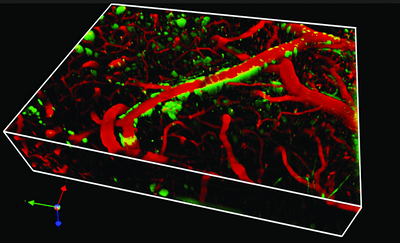
The reason we can only view the this way is, because the vessels in the brain make up a 3D network & the two-photon microscope used in the Schaffer-Nishimura Lab scans it layer by layer. E.g., here is a segment of this network represented in a 3D model:
Except in Stall Catchers we can only look at one later at a time. However, by looking at the consecutive layers with the “virtual microscope” (by “playing” the “vessel movie”) we can get a sense of what the vessel looks like, what is it’s 3D shape, how is it crossing the plane of view, and of course - what’s going on inside it.
The Facebook video shows exactly what you pointed out - at any one time we are looking at a cross-section of the tissue, and depending on the 3D shape of the vessel & how it crosses the plane of view, it may look very different.
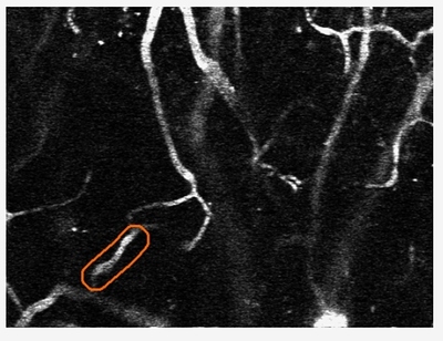
E.g. here’s a vessel seen in it’s entire length in one particular layer, since it’s position in the tissue matches the plane of view:
But others may be vertical to the plane of view or cross it in several points, as in the Facebook video. Here’s another example of a differently shaped vessel & how we see it in the movie: 3D model of Alzheimer's blood vessel | Exclusive look into #Alzheimers brain! See a 3D model of blood capilaries in live mouse brain. Researchers need to analyze thousands of such tiny vessels... | By Human Computation Institute - Facebook
In any case, during the duration of the movie, you should be able to follow the entire vessel within the outline as different parts (layers) of it come in & out of view, and focus on the cross-sections that you see at any one time to determine if there are any stalls. Sometimes that’s easier than other times!
That is true only for the calibration movies (the ones that give you an immediate answer & a high score), as those are the only ones previously seen by experts. The rest is totally up to you guys! (See below why we can be confident in crowd answers.)
The red dots on real movies are those places there by other catchers, which ideally creates a learning opportunity to see how other have interpreted the movie. Albeit that means that there can be mistakes too! (Again, see below for explanation why that doesn’t disturb the overall research.)
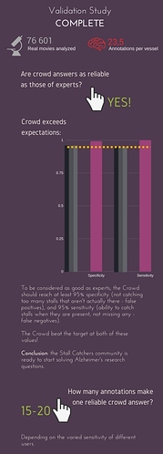
@pietro would be the absolute best person to answer this, as he is the one conjuring the crowd science that makes Stall Catchers work ![]() Hopefully, he will chip in too, but in short: we collect multiple answers per vessel to make sure that the “crowd answer” is correct. We have tested this in our initial validation study last year, and it turned out that we need to collect 15-20 answer to be sure that the crowd answer is as accurate as that of experts (you crowd are GOOD at this!). Quick infographic explains this (click on it for full size):
Hopefully, he will chip in too, but in short: we collect multiple answers per vessel to make sure that the “crowd answer” is correct. We have tested this in our initial validation study last year, and it turned out that we need to collect 15-20 answer to be sure that the crowd answer is as accurate as that of experts (you crowd are GOOD at this!). Quick infographic explains this (click on it for full size):
In other words, this makes sure that we can be >95% confident (and that’s enough for the purpose of the research) that the collective answers of the crowd are correct. The experts will only ever look at the questionable movies, i.e. the one’s where no clear “stalled” or “flowing” answer could be determined by the crowd.
However, we have some exciting developments in this front too! @pietro has been developing new dynamic consensus methods, which make sure that we take each catcher’s sensitivity into account. Pietro has just presented these findings in a talk at the citizen science conference we all went to, and it is definitely worth checking out here: Dynamic Consensus Methods of Stall Catchers (talk at CitSci2017 conference) - YouTube
So, exactly as you pointed out - in theory we could have 10 answers by less experienced users for one movie and thus get a less reliable end result, but then again, we could have 3 answers by our super users for one movie and be extremely confident in their collective answer. The new methods for determining crowd answers will take that into account, measure each catcher’s sensitivity and stop collecting answers for any movie as soon as there’s enough expert-like answers for it. This way we can reduce the average number of annotations needed per vessel to 4-7, which - obviously - makes best use of YOUR time, makes sure everyone’s contributions matter & allow us to analyse data at even greater speeds!
Please let us know if you have any more concerns about this - more on the new methods to appear on the blog soon!
Good point, although both blood clot & stall can eventually loosen up & migrate, in principle. So could a piece of fruit if it is soft enough, for example! But the bottom line here is that something solid (a blood cell) is causing the “straw” (the vessel) to become clogged for an certain amount of time ![]()
That’s what YOU are helping us find out! ![]() read on …
read on …
[quote]Do these stalls in some way cause Alzheimer’s disease, or is there some third variable that is causing both Alzheimer’s disease, and the stalled blood vessels? I’m just curious as to why it’s important to identify these blood vessels as being installed, or flowing. Which hypothesis are you trying to disprove?
[/quote]
This question would be best answered by @nn62, Chris_Schaffer & the rest of the S-N Lab, but in principle the questions you ask is exactly what we are trying to answer with this research! In other words, the researchers at S-N Lab think there’s a link between stalls & Alzheimer’s disease, and that by preventing or reversing stalls you could prevent Alzheimer’s or even reduce existing symptoms - in fact, much of this has already been demonstrated in the lab. Many question marks remain, however, like the exact nature of this link & what molecular pathways we could tackle to find a treatment.
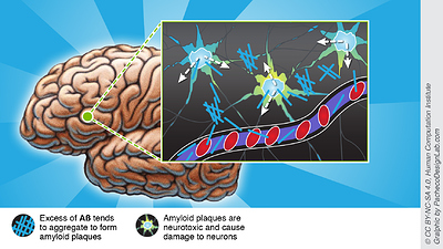
For example, the current dataset is looking at the spatial relationship between stalls & amyloid plaques - if they occur closer together than flowing vessels, that could indicate that one is driving the development of the other, and by targeting & disrupting this destructive feedback loop we could prevent stalls & amyloid forming in the first place, or reduce the symptoms for existing Alzheimer’s patients. As you may know, amyloid plaques are thought to be one of the most likely culprits behind Alzheimer’s disease, as they are neurotoxic & damage the nervous system.
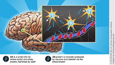
Here’s a little more science in the form of infographics we made previously, which explain why the researchers are interested in stalls & how they could be linked to Alzheimer’s:
What is amyloid beta protein:
How amyloid plaques are formed:
How amyloid plaques could be linked to stalls (and stalls to amyloid plaques):
<img src=“/uploads/default/original/1X/c23c2d67f205b07f84db014e8c23de2e901519c6.jpg” width=“400"”>
Also, you may find this article of mine interesting - it explains the science behind Stall Catchers: http://blogs.discovermagazine.com/citizen-science-salon/2016/04/22/science-behind-wecurealz-citizen-science-alzheimers/
Haha, don’t worry about it - and keep them coming !!!
